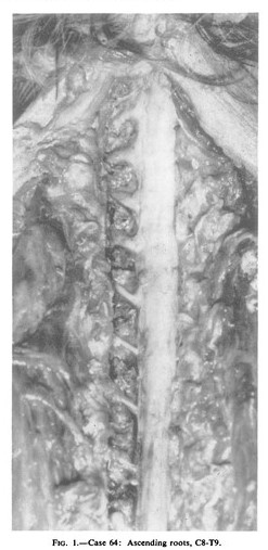
Ascending thoracic nerve roots
Most of us have a textbook view of the nerve roots emerging from the spinal cord and running transversely in the neck and the subsequent spinal nerves (thoracic, lumbar) progressively slanting more vertically. Why? Well, the spinal cord fills the entire spinal canal for the first few months of foetal life. The cord is anchored cranially and at about 4 months the spine begins to grows faster than the cord and thus roots begin to run obliquely.
However, in around 70-75% of people, the thoracic (especially upper) and lower cervical roots ascend in the spinal canal. The percentage of people with ascending roots increasing over the age of 25 years. The anomaly (though we could argue if it is an anomaly) appears to develop in early adult life with no increase in frequency in older groups.
This research comes mainly from dissections of around 130 cadavers by two groups – one in New Zealand (Reid 1960) and in Israel (Nathan & Feuerstein 1970).
Ascending thoracic roots on the treatment couch?
So – my patient is a 22 year old with 13 years of shifting pains, on/off, seemingly growth spurt related and causing marked activity limitations. In the slump long sit position with the neck flexed, adding a little neck lateral flexion is limited and burns sharply around the upper scapula and instantly relieved by a few degrees of knee flexion. I ponder ‘Has this got anything to do with ascending thoracic roots?’
Later on I checked the net – no new research since 1970 – researchers have fled to the brain! So I dug out my old references (from my musty filing cabinet of course – the internet never goes back far enough to the good stuff). No specific causes for the ascending roots could be found – the pathological correlation work was never done and the authors leave it as multiple causes including local dural contraction and adhesion and suggestions that the dura and cord have not “kept up” with spinal canal changes. Growth spurt changes may be involved, yet the anomalies seem more common after the growth spurt. I wondered about general lumbar and lower thoracic spinal inflexibility especially with a western lifestyle – our chairs, our limited squatting, our fear of the lumbar region – is it just pulling the dura down – that’s what it looks like in the image. The two groups both suggest, that the anomalies may make an individual more susceptible to local traumatic lesions and circulatory disturbance. Maybe that is what I am seeing in my patient?
So what?
Well… I would never say ‘wow – I reckon your nerve roots could be angulated and going the wrong way’. But I will continue with movement and educational therapies aiming to liberate whole body movement and function and that also seeks a mechanically permissible spine, meninges and cord, all in a framework with perhaps just a little more understanding of why something hurts.
– David Butler
References
- Reid JD (1960) Ascending nerve roots. J Neurol Neurosurg Psychiatr 23: 148
- Nathan H, Feuerstein M (1970) Angulated course of spinal nerve roots. J Neurosurg 32: 349
And as in many of these neuroanatomy stories, the observations were first made 100 years prior …
- Baldwin WM (1908) The topography of spinal nerve roots. Anat Rec 2:155–156

Melbourne, 31 March – 2 April EP + GMI SOLD OUT
Adelaide, 26-28 May EP + GMI
Wollongong, 14-16 July EP + GMI
Darwin, 4-6 August EP + GMI
Brisbane, 25-27 August EP + GMI
Newcastle, 8-10 September EP + GMI
Hi David,
Really interesting, thanks.
As you suggest 70-75% prevalence probably represents a norm rather than an exception?
Also, what are your thoughts on how this relates to the ‘C6-T6-L4 zones of relative neuro-meningeal immobility’?
Lastly, the image is interesting as the lower thoracic nerve roots pictured pictured (to T9 as described), may perhaps be expected to be more horizontal if the dura -cord were being pulled up to T6?
Max
Hi Max,
It may well relate to the neuromeningeal structures moving downwards in relation to the spinal canal in the zone between C6 and T6 as it adapts from spinal extension to flexion – this feature was demonstrated a number of times in the 80s. I thought that maybe with lumbar (or cervical stiffness) that this feature may have been enhanced but in Reid’s dissections about half of those with ascending roots had tight dura mater and the other half had slack dura.
Thoracic pain is really troublesome – I often think that these tantalising clues to something special going on in the area need more investigation.
Cheers
David
It would be great if the cadaver study were replicated/updated, and compared to other mammals with spines. (Maybe it’s just a developmental phenomenon.)
Hi Diane,
It may well be developmental and comparison with other mammals would be a sensible place to restart research. Some of the neuromeningeal movement studies were carried out on monkeys in the 1950s and 60s and it wasn’t recorded then although it probably wasn’t examined.
All the best
David
Did you consider anomalous root anatomy?
https://books.google.nl/books?id=81meDAAAQBAJ&pg=PA106&dq=anomalous+root+anatomy+Benzel%27s+Spine+Surgery:+Techniques,+Complication+Avoidance,+and+Management&hl=nl&sa=X&ved=0ahUKEwjX4aK7nN7SAhWDVxQKHZA6Az8Q6AEIHDAA#v=onepage&q=anomalous%20root%20anatomy%20Benzel%27s%20Spine%20Surgery%3A%20Techniques%2C%20Complication%20Avoidance%2C%20and%20Management&f=false
page 106
Thanks Marcel,
Anomalies are common in the nervous system – I often wonder when does an anomaly become normal anatomy. These anomalous roots appear to be associated with development early in adult life. There are a lot of unanswered questions though.
Cheers
David